

Published by Zhuhai Biori Biotechnology Co., Ltd.
HCP Risk Perception Series, Part I
What Are Host Cell Proteins?
Host cell proteins (HCPs) are encoded by the host cell’s own genome and expressed as metabolic enzymes, molecular chaperones, secreted proteins, and the like. During cell culture, HCPs enter the harvest cell culture fluid (HCCF) either by active secretion or through release following cell death and lysis (apoptosis with secondary necrosis, or necrosis), and are carried with the target product into downstream purification. Certain HCPs are difficult to remove because they interact with the product or closely resemble it in physicochemical properties, creating a risk of co-purification and carryover into the drug substance.
HCPs are among the most critical process-related impurities in biopharmaceutical manufacturing, and their residual levels bear directly on product safety and efficacy. HCPs exert four principal impacts on biotherapeutics. Immunogenicity: HCPs are non-self proteins that can elicit anti-HCP antibodies even when total HCP is within specification; PLBL2 induced antibodies in most subjects in a clinical study, and clusterin, glutathione S-transferase and peroxiredoxins are also reported. Product quality: host proteases such as HTRA1 and cathepsins B/D cleave or modify the product; PDI and BiP/GRP78 promote aggregation. Formulation stability: lipases and esterases including LPL hydrolyze polysorbate and shorten shelf life, with released fatty acids nucleating particles. Direct biological function: CHO-derived MCP-1/CCL2 retains cytokine activity, and latent TGF-β1 becomes biologically active upon activation in human cells.
For a long time, the industry has relied on ELISA for the total quantitation of HCPs, using “ng/mg” or “ppm” as a single quality control metric[1][2]. However, as understanding of the biological functions of HCPs has deepened, a growing body of evidence demonstrates that the risk posed by an HCP is not equivalent to its total mass—proteases present at trace levels may degrade the product, proteins with adjuvant activity may trigger adverse reactions, and certain highly abundant structural proteins carry almost no safety risk.
This shift in understanding is driving the transition of HCP quality control from “total mass monitoring” to “risk stratification.” In 2021, the BioPhorum Development Group (BPDG) HCP Workstream published a consensus paper, based on a survey of 26 companies (18 of which responded), proposing a four-category framework for classifying high-risk HCPs[3]; USP General Chapter <1132.1>, which became official on May 1, 2025, added informational guidance on mass spectrometry-based HCP testing and thereby provided a methodological basis for risk identification at the single-protein level[4].
This series outlines the upgrade path of the HCP quality control system in four parts, beginning with the evolution of HCP risk perception — from the limitations of total-HCP monitoring to the BPDG four-category risk classification framework. Part 2 analyzes six root causes of inter-assay discrepancies in HCP ELISA results; using quantitative models, it shows how a mismatch between the HCP composition of the calibrator and that of the process sample introduces systematic assay bias, and proposes process-matched and host-matched (single-cell-line–derived) calibrator strategies. Part 3 covers HCP assay selection across the product lifecycle, including stage-specific method choices, a six-step framework for screening candidate methods, and risk assessment of, and regulatory pathways for, method changes. Part 4 addresses the combined use of orthogonal methods and recent regulatory developments, including antibody coverage analysis, the expanding role of LC-MS as an orthogonal method under USP <1132.1>, and targeted control strategies for high-risk HCP species.
Evolution of HCP Risk Perception: From “Total Mass” to “Individual Proteins”
2.1 Limitations of Traditional Total Mass Monitoring
The core logic of traditional HCP quality control is this: assuming that all HCPs carry equal risk, keeping the total below a defined threshold is sufficient to ensure safety. In practice, this logic faces three fundamental challenges:
First, the high heterogeneity of HCP composition. A typical CHO cell harvest can yield thousands of distinct HCPs, with abundances spanning several orders of magnitude[2]. Removal efficiency during purification varies widely among HCPs—ion-exchange chromatography may selectively enrich specific HCPs whose charge properties resemble those of the product, while affinity chromatography may allow certain chaperone proteins to “hitchhike” through co-purification[2][3]. Consequently, the HCP composition remaining in the drug substance differs substantially from that in the harvest: downstream purification imposes strong selective pressure. Readily removable abundant proteins are largely cleared, whereas co-purifying proteins with physicochemical properties close to those of the product are relatively enriched. Commercial kits commonly use harvest material to prepare the calibrator—this is standard industry practice, and it satisfies the needs of process optimization and trend monitoring when process matching is adequate. However, when the calibrator differs substantially from the actual samples in cell line, culture process, or purification flow, mismatches in protein composition and antibody reactivity introduce systematic bias, causing reported values to deviate from the true residual level.
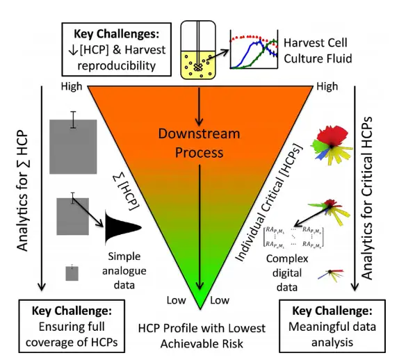
Second, the risk levels of different HCPs differ dramatically. A representative case is phospholipase B-like protein 2 (PLBL2): in some monoclonal antibody products, PLBL2 accounts for less than 5% of total HCP, but because of its high immunogenicity and co-purification with the product, it was classified by BPDG as a high-risk HCP and has become a focus of industry and regulatory attention[3]. Conversely, some highly abundant cytoskeletal proteins (such as actin and tubulin) constitute a large share of total HCP, yet typically have no immunogenicity or enzymatic activity, so their practical risk is limited [2][6].
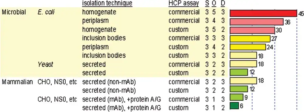
Third, ELISA total-HCP data cannot reflect single-protein risk. ELISA yields a single aggregate value and cannot tell the user which specific proteins are present, what proportion each contributes, or whether known high-risk factors are included[1][2]. When two products both have a total HCP of 10 ng/mg—one dominated by PLBL2, the other dominated by inactive structural proteins—their safety risks are entirely different, yet ELISA cannot distinguish them.
2.2 High-Risk Host Cell Proteins: The BPDG Risk Categories
In 2021, Jones et al. reported a multi-company collaborative study in Biotechnology and Bioengineering that systematically mapped 25 groups of high-risk HCPs in the CHO expression system and classified them into four categories according to their risk mechanism:
| Risk Category | Core Mechanism | Typical Representatives |
|---|---|---|
| Product quality / degradation risk | Enzymatically active; able to directly degrade the product or affect product stability | Cathepsins B/D/L/E/Z; Matrix metalloproteinase (MMP); Alpha-enolase (ENO1); Serine protease (HTRA1); Protein disulfide isomerase (PDI); Peptidyl-prolyl cis-trans isomerase (PPIA); 78 kDa glucose-regulated protein (GRP78/BiP); Carboxypeptidase D (Cpd) |
| Formulation stability risk | Interacts with formulation excipients and affects product shelf life | Sialate O-acetylesterase (SIAE); Carboxylesterase (CEB); Lipoprotein lipase (LPL); Lysosomal acid lipase (LAL); Lysosomal phospholipase A2 (LPLA2) |
| Biological function risk | Cytokine, growth factor, or signaling activity that may interfere with efficacy | Monocyte chemoattractant protein-1 (MCP-1/CCL2); transforming growth factor β (TGF-β); C-X-C motif chemokine 3 (CXCL3) |
| Immunogenicity risk | Adjuvant activity or high immunogenicity; may induce immune responses in patients | Clusterin (CLU); Annexin A5 (ANXA5); Glutathione-S-transferase (GST); Peroxiredoxin (PRDX); Phospholipase B-like 2 (PLBL2); S100 protein family; Procollagen-lysine 2-oxoglutarate 5-dioxygenase 1 (PLOD1); Pyruvate kinase (PK) |
The paper also notes that a subset of HCPs is difficult to remove primarily because they co-purify with the product—either through direct, typically non-specific interaction with the product molecule (hydrophobic interactions, hydrogen bonding, van der Waals forces, and ionic interactions), or because their physicochemical properties (pI/charge, hydrophobicity, molecular weight) resemble those of the product. Although such proteins may lack direct toxicity or immunogenicity, they are poorly cleared by conventional platform chromatography steps, and persistent species can account for a substantial fraction of the residual HCP in the drug substance[2]. Typical representatives include abundant, functionally inert proteins repeatedly detected after Protein A capture—such as elongation factor 1-alpha (eEF1a) — as well as ribosomal proteins (e.g., 40S ribosomal protein SA, RPSA) listed among the frequently observed CHO HCPs[3]. Protein disulfide isomerase (PDI, PDIA1; gene symbol P4HB) is also frequently cited as difficult to remove, although the BioPhorum framework assigns it an aggregation impact.
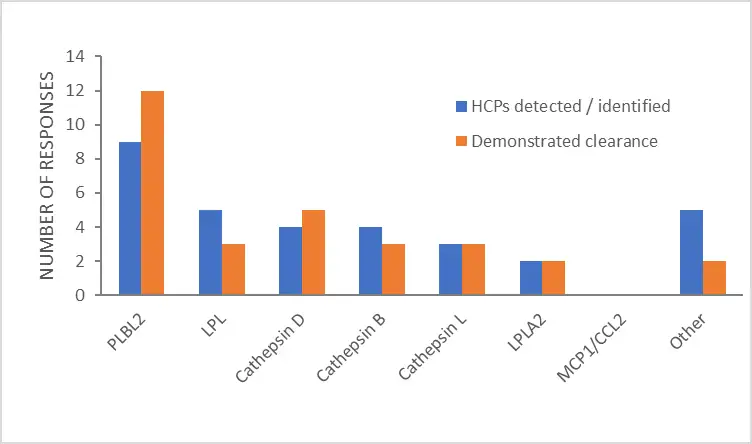
2.3 Differences in High-Risk HCPs Across Expression Systems
The high-risk HCP profile is strongly expression-system specific. Applying the CHO high-risk list directly to E. coli, HEK293, or other expression systems may result in missing critical risks. Below is a comparison of high-risk HCP characteristics across the major expression systems.
E. coli expression system: Unlike mammalian cells, the most prominent risk among E. coli HCPs is immunogenicity. Flagellin (FliC) acts as a potent TLR5 ligand; even trace residues may trigger a strong innate immune response[6]. Heat shock proteins (DnaK, GroEL, IbpB) and outer membrane proteins (OmpA, OmpF) are also predicted to carry immunogenicity risk[6]. In addition, endogenous E. coli proteases—Lon, DegP, OmpT and Ptr (protease III)—can degrade recombinant protein products, particularly after cell lysis[7,8]. Notably, significant differences exist among strains: BL21 strains are deficient in the Lon and OmpT proteases, whereas K-12 strains such as W3110 retain both[21].
HEK293 expression system: In gene therapy and vaccine products manufactured in HEK293 cells, the residual HCP profile reflects both the host cell proteome and the stresses imposed by transient transfection and rAAV production. Caspases-2, -3, -7, and -8 were among the differentially expressed proteins associated with apoptosis in AAV-producing HEK293 cells[9], consistent with the caspase-mediated cell death observed during rAAV production[10]. Proteomic profiling of AAVX affinity eluates identified the heat shock proteins HSPA1B, ST13, and HSP90AB1 as the three highest-abundance residual HCPs, all involved in binding misfolded or aggregated protein assemblies[11]. Nucleic acid-binding proteins such as nucleolin (NCL) and nucleophosmin (NPM1) may associate with AAV particles[12,13]. LGALS3BP (90K) has been shown to co-purify with AAV6[14] and was identified as the sole major HCP contaminant in clinical-grade AAV8 lots[15]. Several of these persistent HCPs—alpha-enolase and PPIA (drug quality), BiP/HSPA5 (drug quality, aggregation), and pyruvate kinase and peroxiredoxin-1 (immunogenicity)—fall within the high-risk categories of the BPDG framework[11]. Immunoinformatic tools such as ISPRI-HCP have been applied to classify process-related impurities derived from HEK293-based manufacture according to immunogenicity risk [6].
MDCK expression system: As one of the primary production cell lines for influenza vaccines, MDCK cells present a set of high-risk HCP candidates with canine-specific characteristics: GSTM4, the most abundant detectable phase II metabolic enzyme (low in human tissues); ATP1A1 (Na⁺/K⁺-ATPase α1), among the most abundant protein species in MDCK cells; and CYP4A47, a cytochrome P450 fatty acid hydroxylase highly expressed in the canine kidney[16]. Process-level evidence confirms that ANXA2, ACTG, and PRDX1 persist throughout MDCK-based vaccine purification[17]. In addition, trypsin supplementation—required to cleave the hemagglutinin precursor (HA0) and activate viral infectivity—is not a canine HCP and must be monitored separately as a process-related impurity.
Sf9 insect cell expression system: The HCP risks of the baculovirus–insect cell expression system are distinctive. Sf-caspase-1 (P89116), the effector caspase orthologous to human caspases-3 and -7 and the target of the baculoviral anti-apoptotic protein p35, is activated and released in large amounts during late-stage baculovirus infection[18]. Sf-cathepsin L is among the best-characterized endogenous Sf9 proteases. Because insect proteins often show limited sequence identity with their human orthologs, immunogenicity is the most prominent risk dimension of the Sf9 system. In addition, proteins encoded by the baculovirus itself—including the envelope glycoprotein GP64 (P17501), the viral cathepsin V-CATH (P25783), and the chitinase CHIA (P41684)—must be included in the risk assessment as key process-related impurities[19]. Another specific challenge of the Sf9 system is that cell viability at harvest typically ranges from 50% to 70% at the optimal time of harvest, and lysis of a substantial cell fraction releases large amounts of intracellular proteins, resulting in a high HCP burden[20].
From Classification to Assessment: A Clinical Safety Risk Assessment Framework for High-Risk HCPs
The impact-based classification of high-risk HCPs has recently been extended from identification to formal clinical safety risk assessment. In an updated industry review, the BioPhorum Development Group (BPDG) HCP Workstream surveyed current practice and found that companies diverge in when they identify individual HCPs by LC-MS/MS: only 1% do so routinely before IND, 18% routinely after IND, 30% only for root cause analysis, and 48% use a combined approach—identification typically being triggered by elevated total HCP levels, inconsistent process performance, product stability issues, or dilutional nonlinearity in the HCP ELISA [21].
Once an individual HCP has been identified and quantitated, the recommended practice is to conduct a clinical risk assessment in parallel with the product quality risk assessment. The framework—developed from the earlier de Zafra model—comprises two parallel evaluations, for biological activity and for immunogenicity, in which HCP-related factors (known function, HCP dose derived from the ng/mg level and the therapeutic dose, homology to the human orthologue, and accessibility of that orthologue at its site of action) are weighed against clinical factors (indication, patient population, mechanism of action, route, frequency and duration of administration, and prior experience with the HCP). The weighting is deliberately case-by-case rather than a numerical scoring tool.
Supporting data accumulate across the development lifecycle: prior knowledge—literature, internal databases, and the BPDG watchlist of high-risk HCPs—is combined with in silico tools such as CHOPPI, IEDB, EpiVax, ISPRI-HCP, and JanusMatrix[6,19], in vitro assays based on PBMCs or dendritic cells, and nonclinical toxicology, while recognizing that clinical data remain the ultimate arbiter and that animal studies conducted solely to characterize HCP risk are discouraged. Notably, total HCP level does not translate directly into immunogenic risk: in vitro, total HCP up to 4,000 ppm did not augment immune responses [21].
The PLBL2 case study illustrates the discriminating power of this framework: the same impurity was judged moderate-to-high risk in allergic asthma (subcutaneous, chronic dosing) but low-to-moderate in oncology (intravenous, weekly dosing in an immunosuppressed population). Although patients in the originating Phase 3 study developed anti-PLBL2 antibodies, no adjuvant effect or safety signals were observed [21]. Consistent with the ALARA principle, moderate-to-high risk outcomes should drive process improvement, supported by cross-functional teams spanning CMC, nonclinical, and clinical functions [21].
Two Lines of Defense: Process Clearance and the Analytical Toolbox in HCP Control Strategy
The core of an HCP control strategy is to treat process clearance as the first line of defense and the analytical toolbox as the second—each verifying the other—rather than relying on a single release test.
Process side (first line). The HCP profile is largely determined upstream: cell line and clone selection, medium formulation, culture duration, and harvest conditions shape which proteins are expressed, released, and degraded; titers gained at the cost of harvest viability increase cell lysis. Because a subset of HCPs hitchhikes through the process—via non-specific interactions with the product or resin, physicochemical similarity to the product, or sheer abundance in HCCF—upstream decisions predetermine which species will be difficult to remove. The process logic is therefore to assign clearance tasks by HCP type: abundant, soluble HCPs with physicochemical properties distant from the product are cleared mainly by routine operations such as deep filtration, low-pH hold, and anion exchange flow-through; co-purifying proteins—high-pI ribosomal proteins and elongation factors, and chaperones binding the molecule non-specifically through hydrophobic contacts or hydrogen bonds—require targeted process design, such as high-salt elution, detergent or arginine washes, mixed-mode chromatography, or adjusted wash conditions around Protein A; HCCF acidification further reduces co-purification of high-risk species. Evidence of clearance comes from stage-wise HCP profiles collected and compared across steps, not from the residual total of the final batch alone; under the ALARA principle, early-stage total HCP specifications for mAbs are typically set around 100 ng/mg and tightened based on process capability and clinical experience[21].
Analytical side (second line). The analytical toolbox must combine abundance with identity. The multi-analyte HCP ELISA governs release decisions and batch consistency, quantified against a process-matched calibrator—but it reports only total ppm, covers only the antigens the immunizing animal recognizes, and is not comparable between assays. LC-MS/MS per USP General Chapter <1132.1> (or an equivalent orthogonal method) is therefore required to provide identity, relative abundance, and clearance trajectories: confirming that high-risk watchlist members have indeed been cleared and identifying species missed by the ELISA; once a specific HCP has been characterized, a single-analyte ELISA serves as a quantitative tool for process monitoring or safety specifications [21]. The combination serves two purposes simultaneously: a comparable quantitative baseline for release, and individualized evidence for risk assessment—when total levels or process consistency deviate, identification data feed the biological-activity and immunogenicity assessments[21].
Together, they upgrade “total HCP within specification” to “critical HCPs under control”: orthogonal methods applied across the lifecycle—from HCCF characterization to drug substance release—turn the control strategy from a static specification into a self-correcting system in which identification data continuously feed back into process optimization, closing the clearance–analysis loop[2].
Chapter Summary
The shift from “total mass monitoring” to “risk stratification” is not merely an upgrade of the HCP quality-control concept; it also deepens our understanding of safety risks in biopharmaceutical products. Traditional total mass monitoring assumes that all HCPs carry equal risk, whereas the risk stratification concept recognises that HCP composition is highly heterogeneous, that risk levels differ dramatically among HCPs, and that ELISA total-HCP data cannot reflect single-protein risk. The BPDG four-category risk classification framework provides a systematic tool for identifying high-risk HCPs; and from classification to assessment, the clinical safety risk assessment framework takes this one step further — weighing the biological activity, dose-normalised exposure, human homology, route of administration and patient population of each residual HCP to judge, case by case, whether it matters clinically. Meanwhile, the differences in high-risk HCP profiles across expression systems remind us that there is no “universal” HCP testing method: selection must be tailored to the specific expression system and process characteristics of the product. In practice, this understanding is embodied in two lines of defense — process clearance and the analytical toolbox — in which unit operations are tasked with clearing defined classes of HCPs, while a combined analytical strategy of total-HCP ELISA plus orthogonal methods verifies the clearance and exposes what it misses. Classification tells us which HCPs deserve attention; assessment tells us how much they matter; and the two lines of defense prove that they are under control.
In the Next Installment
In the next article, we will explore in depth the six root causes of variability in ELISA reporting, use a quantitative model to analyze the impact of mismatched calibrator composition on quantitation results, and detail the principle of process matching and the strategy for selecting single-cell-line calibrators.
FAQ
Can trace HCPs damage a drug during storage?
Yes. Although present at trace levels, certain HCPs can degrade a biopharmaceutical product during storage, because the relevant attribute is enzymatic activity rather than abundance — a catalyst acts turnover-wise, so picomolar amounts can accumulate substantial damage over shelf life. The best-documented pathway is polysorbate degradation: host-cell lipases and esterases — lipoprotein lipase (LPL), lysosomal acid lipase, lysosomal phospholipase A2 and sialate O-acetylesterase — hydrolyse polysorbate 20/80, and the released free fatty acids bind to the antibody through hydrophobic interaction and drive particle formation, shortening formulation shelf life. Direct evidence comes from a 2017 CHO study in which knockout of the endogenous LPL gene markedly reduced polysorbate degradation, confirming LPL as a principal degrading enzyme and showing that identity-targeted removal can be more effective than lowering the total HCP burden. Host proteases (cathepsins B, L and D, HTRA1) can likewise cleave or modify the product, and such activity persists throughout the product life cycle, including during storage. Importantly, identity matters more than abundance: PLBL2, once assumed to degrade polysorbate, was later shown in vitro to have no hydrolase activity. Storage-phase risk should therefore be verified through accelerated stability together with polysorbate content, free fatty acid and particle analysis, and where relevant an enzymatic activity assay — not by total HCP alone.
Are serum proteins and added enzymes counted as HCPs?
No, not in the strict sense. Under ICH Q6B, HCPs are proteins derived from the production host cell; serum proteins (e.g., BSA or other foetal bovine serum components) are medium-derived impurities, and intentionally added enzymes (trypsin, DNase/Benzonase, lysozyme) are exogenous processing reagents. All three belong to the broader class of process-related impurities, so all must be controlled, but they should be classified, monitored and reported separately rather than merged into a single HCP figure. A practical caveat is assay cross-reactivity: the polyclonal antibodies used in multi-analyte HCP ELISA can recognise medium-derived proteins or added enzymes, which inflates the reported HCP value and may mask the true host-cell protein profile; for processes using serum-supplemented media or added enzymes, cross-reactivity should be assessed, or chemically defined media adopted, with added-enzyme residues measured by a dedicated assay.
Does a higher HCP result always mean a higher clinical risk?
No. A total HCP value is a measure of exposure, not of hazard. Under ICH Q6B, acceptance criteria should be established from process capability together with product characterisation and nonclinical/clinical data, which means the clinical risk of a given HCP depends on three things: its identity and biological activity (whether it is on the watchlist, retains enzymatic or immunomodulatory function, or is homologous to a human protein), its dose-normalised exposure (ng/mg multiplied by the therapeutic dose in mg/kg), and the available clinical experience in the relevant indication and patient population. The literature contains clear counter-examples in both directions: PLBL2 in lebrikizumab was present at only 242–328 ng/mg yet induced anti-PLBL2 antibodies in 76%–90% of subjects, whereas CHO HCP in trenonacog alfa reached 58,500 ng/mg with 29% anti-HCP antibody incidence and no associated adverse events. The widely used 100 ng/mg limit is a historical industry benchmark, not a scientifically derived threshold, so a result above it is a trigger for investigation and risk assessment rather than an automatic safety signal.
References
- USP General Chapter <1132> Residual Host Cell Protein Measurement in Biopharmaceuticals.
- Wang X, Hunter AK, Mozier NM. Host cell proteins in biologics development. Biotechnol Bioeng. 2009;103(3):446-458.
- Jones M, Palackal N, Wang F, Gaza-Bulseco G, Hurkmans K, et al. “High-risk” host cell proteins (HCPs): A multi-company collaborative view. Biotechnol Bioeng. 2021;118(8):2870-2885.
- USP General Chapter <1132.1> Residual Host Cell Protein Measurement in Biopharmaceuticals by Mass Spectrometry. USP-NF 2025.
- Bracewell DG, Francis R, Smales CM. The future of host cell protein (HCP) identification during process development and manufacturing linked to a risk-based management for their control. Biotechnol Bioeng. 2015;112(9):1727-1737. doi:10.1002/bit.25628.
- Haltaufderhyde K, Roberts BJ, Khan S, Terry F, Boyle CM, McAllister M, Martin W, Rosenberg A, De Groot AS. Immunoinformatic Risk Assessment of Host Cell Proteins During Process Development for Biologic Therapeutics. AAPS J. 2023;25(5):87.
- Rosano GL, Ceccarelli EA. Recombinant protein expression in Escherichia coli: advances and challenges. Front Microbiol. 2014;5:172. doi:10.3389/fmicb.2014.00172.
- Karyolaimos A, de Gier J-W. Strategies to enhance periplasmic recombinant protein production yields in Escherichia coli. Front Bioeng Biotechnol. 2021;9:797334. doi:10.3389/fbioe.2021.797334.
- Strasser L, Boi S, Guapo F, Donohue N, Barron N, Rainbow-Fletcher A, Bones J. Proteomic landscape of adeno-associated virus (AAV)-producing HEK293 cells. Int J Mol Sci. 2021;22(21):11499.
- Ladiwala P, Ndahiro N, Hauk P, Wen J, Sargunas J, Chen YJ, Barton E, Betenbaugh MJ. Unraveling cytotoxicity in HEK293 cells during recombinant AAV production for gene therapy applications. Biotechnol J. 2025;20(3):e202400501.
- Leibiger TM, Min L, Lee KH. Quantitative proteomic analysis of residual host cell protein retention across adeno-associated virus affinity chromatography. Mol Ther Methods Clin Dev. 2024;32(4):101383.
- Qiu J, Brown KE. A 110-kDa nuclear shuttle protein, nucleolin, specifically binds to adeno-associated virus type 2 (AAV-2) capsid. Virology. 1999 May 1;257(2):373-382.
- Bevington JM, Needham PG, Verrill KC, Collaco RF, Basrur V, Trempe JP. Adeno-associated virus interactions with B23/Nucleophosmin: identification of sub-nucleolar virion regions. Virology. 2007 Jan 5;357(1):102-113.
- Denard J, Jenny C, Blouin V, Moullier P, Svinartchouk F. Different protein composition and functional properties of adeno-associated virus-6 vector manufactured from the culture medium and cell lysates. Mol Ther Methods Clin Dev. 2014;1:14031.
- Seitz IP, et al. Investigating the Immunogenic Potential of Variations in Host Cell Protein Levels in Clinical-Grade AAV8 Products. Invest Ophthalmol Vis Sci. 2025;66(6):38.
- Harwood MD, Zettl K, Weinheimer M, Pilla-Reddy V, Wiśniewski JR, Neuhoff S, et al. Interlaboratory Variability in the Madin-Darby Canine Kidney Cell Proteome. Mol Pharm. 2023;20(7):3505-3518.
- Liu B, Li F, Yang Y, Tu H, Hao P, Ma Q, Le Y, Zhang Q. In-depth monitoring of host cell proteins in influenza vaccines throughout multi-step purification processes. Vaccine. 2026;88. doi:10.1016/j.vaccine.2026.128957
- Ahmad M, Srinivasula SM, Wang L, Litwack G, Fernandes-Alnemri T, Alnemri ES. Spodoptera frugiperda caspase-1, a novel insect death protease that cleaves the nuclear immunophilin FKBP46, is the target of the baculovirus antiapoptotic protein p35. J Biol Chem. 1997;272(3):1421-1424.
- Slack JM, Kuzio J, Faulkner P. Characterization of v-cath, a cathepsin L-like proteinase expressed by the baculovirus Autographa californica multiple nuclear polyhedrosis virus. J Gen Virol. 1995;76(Pt 5):1091-1098.
- Carvalho SB, Silva RJS, Moreira AS, Cunha B, Clemente JJ, Alves PM, Carrondo MJT, Xenopoulos A, Peixoto C. Efficient filtration strategies for the clarification of influenza virus-like particles derived from insect cells. Sep Purif Technol. 2019;218:81-88.
- Coye L, Jones MA, Gaza-Bulseco G, et al. Host Cell Protein Clinical Safety Risk Assessment—An Updated Industry Review. Biotechnol Bioeng. 2025;122:3229-3248.