


Published by Zhuhai Biori Biotechnology Co., Ltd.
Summary. phi29 multiple displacement amplification takes picograms of double-stranded DNA to tens of micrograms in one isothermal incubation at 30 °C, without thermal cycling, with product strands averaging more than 10 kb [1,2]. It works because phi29 displaces the strand ahead of the growing 3′ end and re-primes on it, so amplification branches into a network instead of running in cycles. The limit is uniformity rather than yield: yield stops when the dNTP pool runs out, while coverage is set by priming statistics, branch structure and chimera formation, and at trace input that is the harder problem [6,7].
Key takeaways
- phi29 stays bound for ~70,000 nucleotides per binding event and displaces the strand ahead of it, so MDA needs no helicase, no clamp protein and no temperature cycling [3,4].
- At 1–10 genome copies, MDA yields 20–30 µg with locus-to-locus bias under 3-fold, against 4–6 orders of magnitude for PCR-based WGA [2].
- Bias is reduced, not removed: genome representation reaches 99.82%, yet six regions totalling up to 5.62 Mb failed to amplify, and single-cell skew persists [5,6,7,14].
- Three things break trace-input MDA — amplifiable DNA carried by the enzyme, chimeras from displaced 3′ ends, and coverage skew. Each one is a measurable release spec, not a vague notion of “quality” [8,9].
- BR3P104, 3 lots, 0.1 ng human gDNA, 30 °C × 16 h, 20 µL: 0.92–0.95 µg (BIORI internal data).
How phi29 multiple displacement amplification copies a genome without a helicase
One 66 kDa polypeptide does three jobs at once — it polymerises, it proofreads, and it shoves the downstream strand out of the way, so replication proceeds without the accessory machinery a cellular replisome needs.
phi29 DNA polymerase, a strand displacement DNA polymerase, is the product of gene 2 of Bacillus subtilis phage phi29, a family B enzyme supplied as a recombinant protein expressed in E. coli. Three of its properties matter for whole genome amplification:
- Processivity. It adds an average of ~70,000 nucleotides per binding event, the highest reported for a DNA polymerase working without a processivity factor [3,4].
- Strand displacement. It unwinds the duplex ahead of the growing 3′ end and replicates the entire 19,285 bp phage genome without a helicase [3,4]. In MDA this is the amplification engine: the displaced strand is single-stranded, random hexamers anneal to it, and every new 3′ end starts another branch.
- Proofreading. The intrinsic 3′→5′ exonuclease keeps the error rate of amplified product at 9.5 × 10⁻⁶, the same as in unamplified DNA [5].
The reaction is short to describe and long to incubate:
- Template is denatured (95 °C 3 min, snap-chilled) so hexamers can reach both strands.
- Random hexamers anneal and supply 3′-OH primers. They are exonuclease-resistant (phosphorothioate) in modern formulations, because the enzyme’s own exonuclease would otherwise digest them.
- At 30 °C the polymerase extends, displaces, and re-primes on the displaced strand. What accumulates is a hyperbranched network, not a linear amplicon (Figure 1, panel 4).
- dNTPs are consumed until the pool runs out. Yield is capped by nucleotide supply long before it is capped by template copy number.
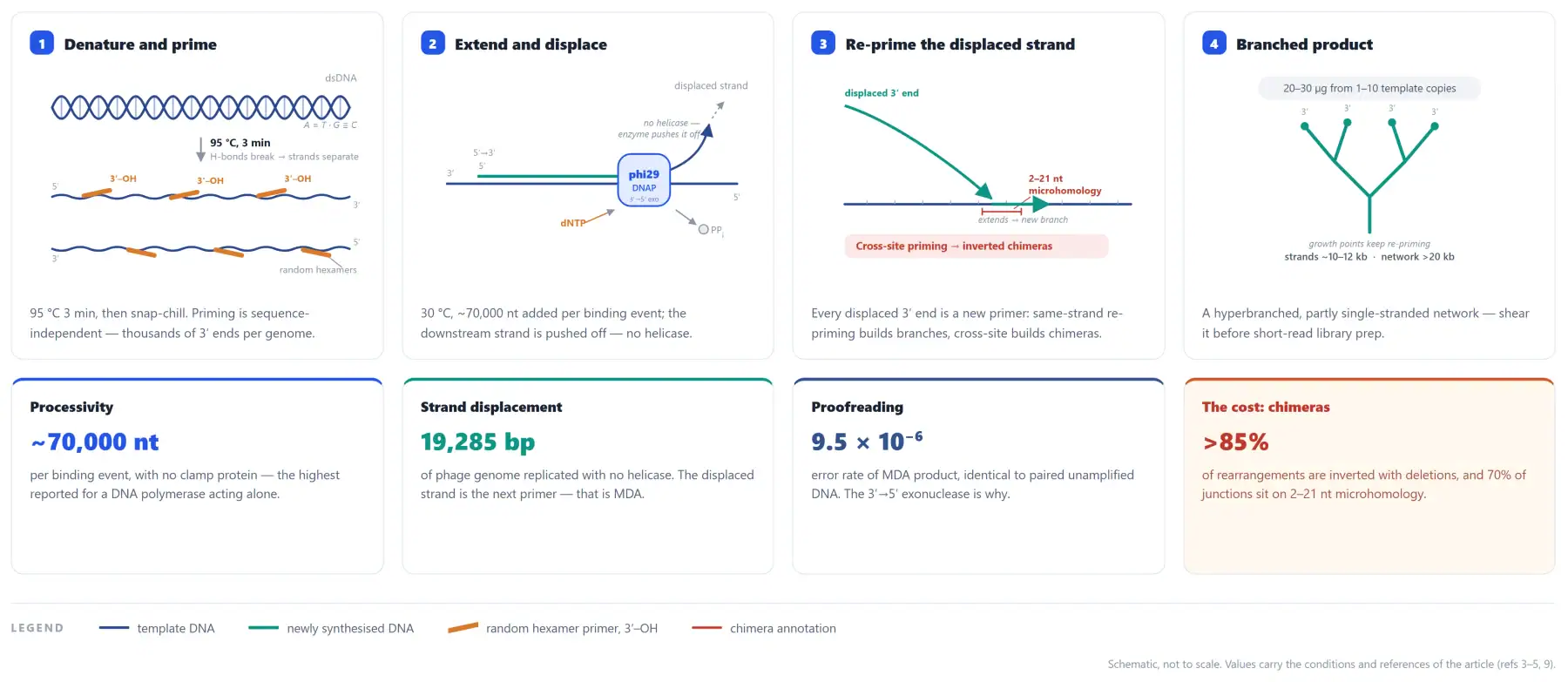
Two consequences follow. Product is a branched, partly single-stranded network that migrates above 20 kb in agarose and has to be fragmented or sheared before short-read library prep. And because the chemistry is isothermal and primer-driven, it will also amplify DNA you never intended to put in the tube — recombinant phi29 purified from E. coli carries host and vector DNA, and it shows up as product in no-template controls [8].
The workflow, and the four parameters that decide it
A working trace-input MDA is five steps, and most failures trace back to template denaturation, primer excess, incubation length, or a missing NTC.
| Step | What to do | Parameter that matters |
|---|---|---|
| 1. Denature | 95 °C 3–5 min → ice 5 min, or alkaline denaturation 5 min at room temperature (cells: alkaline lysis, then neutralise) | Alkaline denaturation is gentler on trace template, but incomplete denaturation under-primes AT-rich and supercoiled regions |
| 2. Assemble on ice | Add enzyme last, keep its volume at 1–2% of the reaction | A large glycerol bolus slows an isothermal reaction and dilutes the Mg²⁺ |
| 3. Extend | 30 °C, 6–16 h, sealed tube | 16 h for ≤100 pg; longer adds chimera as well as yield |
| 4. Inactivate | 65 °C 10 min | Do not skip before quantification or library prep |
| 5. Quantify | Qubit dsDNA HS or PicoGreen | A260 over-reads, because residual dNTP absorbs at 260 nm |
Three knobs the protocol sheet usually leaves at default.
Denaturation chemistry and incubation temperature are copied from whichever kit the lab bought first, and at trace input both change the result more than the enzyme dose does.
- Alkaline denaturation is the gentler option at low input. Alkaline denaturation avoids the damage a 95 °C step can do to trace template and releases supercoiled and AT-rich regions more evenly; we document it as an option rather than a default, because we have not yet quantified the difference on our own lots.
- Temperature belongs to the enzyme. This line is specified at 30 °C. Thermostable engineered variants run at 42 °C and are a different reagent with their own protocol — do not move a wild-type lot to 42 °C because another product runs there [11].
- Time to plateau is set by input. In a real-time-monitored MDA reaction the time needed to reach a given amount of product rose linearly with the log of the input, across the eight tenfold dilution points from 0.1 ng to 0.01 fg — a 7-log range in mass [15], and reviews put the working window at 2–16 h depending on how much template the reaction starts with [16].
Three practical notes that cost people a run:
- EDTA competes for the 10 mM Mg²⁺. Template eluted in TE is usually fine, but a sample carried over from an EDTA-containing lysis buffer will suppress amplification with no visible sign until you sequence the product.
- Primer concentration is fixed at 50 µM whatever the input. At 10 pg that is a >10¹²-fold molar excess over template, so primer-derived synthesis competes with the real template for the same dNTP pool. Adding more primer buys background, not sensitivity.
- Run the NTC every time. In trace work the negative control is the enzyme QC — if the NTC amplifies, the enzyme is your template.
Where MDA is the right tool, and what each application asks of the enzyme
MDA wins wherever template is countable rather than weighable and where product length or locus representation matters more than turn-around time. The corollary: “a good MDA enzyme” means different things in different labs.
Single-cell and low-input sequencing is what moved MDA from a niche trick to a standard method [12,13].
| Method | Mechanism | Thermal cycling | Typical product | Bias profile | Where it wins |
|---|---|---|---|---|---|
| MDA (phi29) | Strand displacement, random hexamer primed | None, 30 °C | >10 kb, branched | Highest coverage below ~10⁶-fold gain and lowest per-base error, but uniformity falls as gain rises [6,14] | Whole genome from a few copies; long-range assembly |
| MALBAC | Quasi-linear pre-amplification, then PCR | Yes | Short | More uniform than MDA and largely insensitive to gain: higher fractional genome coverage above 10⁶-fold gain, and a duplication-resolution window near 3.3 × 10⁴ bp where MDA’s is gain-dependent [10,14]; at a fixed sequencing depth the advantage is partly offset by a larger share of unmappable, primer-derived reads [14] | CNV detection at high gain, where MDA’s bias and CNV resolution worsen as gain rises [14] |
| PicoPLEX / SurePlex | PCR-based, proprietary priming | Yes | Short | Intermediate [7] | Aneuploidy screening, STR profiling |
| DOP-PCR / PEP | Degenerate or random priming, Taq | Yes | Short | Highest — 4–6 orders of magnitude across loci [2] | Legacy workflows, SNP genotyping from ng input |
| Application | Why MDA | What the enzyme must do | Failure mode when it doesn’t |
|---|---|---|---|
| Single-cell and low-input genome sequencing (CTC, uncultured bacteria, blastomeres) | Whole genome from one copy; long product supports assembly | High processivity and activity at ≤10 pg; lowest amplifiable DNA; low chimera | Loci lost at the first priming event; assemblies fragment at chimeric junctions |
| Metagenomics, virome, low-biomass environmental DNA | Bulk DNA often <1 ng; needs community-wide representation | Host and vector DNA carryover below detection; tolerance to humic acids, collagen, EDTA | Enzyme-derived E. coli sequence enters the community profile [8] |
| Clinical CNV, PGT-A, products of conception | Shallow WGS is read on coverage uniformity, not yield | Repeatable lot-to-lot bias pattern; DNase-free; low chimera | Small CNVs sink below the noise floor of representation bias [7] |
| Forensic, FFPE, degraded and ancient DNA | Template is fragmented and lesion-rich; every molecule counts | Tolerates blocked 3′ ends and nicks; high enzyme-to-template ratio; adds no further damage | Lesions stall the polymerase; apparent allelic dropout |
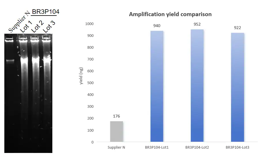
What these numbers do not establish: we have not separated specific activity from processivity; we have not validated FFPE or ancient DNA with these lots. All figures here are 30 °C and 16 h; comparisons with other suppliers hold only when temperature and time are pinned.
FAQ
Must the DNA template be circular for MDA?
No. Circularity was the starting case, not a requirement. The original multiply-primed RCA used plasmid and phage DNA because a single primer can travel round a circle indefinitely, which is how 10,000-fold amplification was reached in a few hours [1]. MDA on linear genomic DNA works differently: random hexamers prime at thousands of positions, and every displaced strand becomes a new priming site. Product from linear template averages >10 kb per strand with genome-wide representation [2,5]. Circular templates (plasmids, phage, circoviruses) are still amplified more efficiently per priming event, which is why plasmid RCA is close to quantitative while genomic MDA plateaus on the dNTP pool.
Does MDA need a thermal cycler?
No. phi29 amplification is isothermal, 30 °C, in a heat block, water bath or incubator; the only other heat steps are the initial denaturation (95 °C, or alkaline at room temperature) and a 10-minute 65 °C inactivation. A thermocycler helps with two optional extras: a ramp-down into the incubation temperature that reduces non-specific priming, and a 4 °C hold before cleanup. On a block, seal the tubes — 16 h at 30 °C loses enough water to change your concentrations.
Does a high MDA yield guarantee uniform genome coverage?
No. Yield saturates on nucleotide supply: 1 mM each dNTP in 50 µL supports roughly 65 µg of dsDNA in theory, and reactions typically plateau at about 20–30 µg regardless of template copy number [2]. Uniformity is set by priming stochasticity, branch structure and chimera formation, and it degrades far faster than yield as input falls. Measure uniformity as its own spec (≥1× coverage, 0.5–2× fraction, depth CV, chimera rate) at a stated input and a stated depth. Then read it as GC-normalized coverage against an unamplified control, rather than from depth CV alone.
About BIORI
Zhuhai Biori Biotechnology Co., Ltd. develops recombinant enzymes and RUO reagent kits for NGS and molecular diagnostics. Recombinant expression, fermentation, purification and formulation run in-house, with lot-to-lot consistency controls at critical process steps and release testing on every lot.
- phi29 DNA Polymerase, BR3P104 (500 U / 1000 U, supplied with 10× phi29 buffer): https://biori-en.com/product/acugenix-phi29-dna-polymerase-br3p104/
- NGS library preparation product selection: https://biori-en.com/product/ngs-selection/
- Manufacturing and lot release: https://biori-en.com/manufacturing-quality/
For custom formulation — glycerol-free, high-concentration, or OEM bulk — contact the team through the site.
For research use only. Not for use in diagnostic procedures.
References
- Dean FB, Nelson JR, Giesler TL, Lasken RS. Rapid amplification of plasmid and phage DNA using phi29 DNA polymerase and multiply-primed rolling circle amplification. Genome Research. 2001;11(6):1095–1099. https://genome.cshlp.org/content/11/6/1095
- Dean FB, Hosono S, Fang L, et al. Comprehensive human genome amplification using multiple displacement amplification. PNAS. 2002;99(8):5261–5266. https://www.pnas.org/content/pnas/99/8/5261.full.pdf
- Blanco L, Bernad A, Lázaro JM, Martin G, Garmendia C, Salas M. Highly efficient DNA synthesis by the phage phi29 DNA polymerase. Symmetrical mode of DNA replication. Journal of Biological Chemistry. 1989;264(15):8935–8940. https://www.sciencedirect.com/science/article/pii/S002192581881883X
- Rodríguez I, Lázaro JM, Blanco L, Kamtekar S, Berman AJ, Wang J, Steitz TA, Salas M, de Vega M. A specific subdomain in phi29 DNA polymerase confers both processivity and strand-displacement capacity. PNAS. 2005;102(18):6407–6412. https://pmc.ncbi.nlm.nih.gov/articles/PMC1088371/
- Paez JG, Lin M, Beroukhim R, et al. Genome coverage and sequence fidelity of phi29 polymerase-based multiple strand displacement whole genome amplification. Nucleic Acids Research. 2004;32(9):e71. https://pubmed.ncbi.nlm.nih.gov/15150323/
- Pinard R, de Winter A, Sarkis GJ, et al. Assessment of whole genome amplification-induced bias through high-throughput, massively parallel whole genome sequencing. BMC Genomics. 2006;7:216. https://pubmed.ncbi.nlm.nih.gov/16928277/
- Deleye L, Tilleman L, Vander Plaetsen AS, Cornelis S, Deforce D, Van Nieuwerburgh F. Performance of four modern whole genome amplification methods for copy number variant detection in single cells. Scientific Reports. 2017;7:3422. https://pubmed.ncbi.nlm.nih.gov/28611458/
- Takahashi H, Yamazaki H, Akanuma S, et al. Preparation of phi29 DNA polymerase free of amplifiable DNA using ethidium monoazide, an ultraviolet-free light-emitting diode lamp and trehalose. PLoS ONE. 2014;9(2):e82624. https://journals.plos.org/plosone/article?id=10.1371/journal.pone.0082624
- Lasken RS, Stockwell TB. Mechanism of chimera formation during the multiple displacement amplification reaction. BMC Biotechnology. 2007;7:19. https://pmc.ncbi.nlm.nih.gov/articles/PMC1855051/
- Chen M, Song P, Zou D, Hu X, Zhao S, Gao S, Ling F. Comparison of multiple displacement amplification (MDA) and multiple annealing and looping-based amplification cycles (MALBAC) in single-cell sequencing. PLoS ONE. 2014;9(12):e114520. https://pmc.ncbi.nlm.nih.gov/articles/PMC4259343/
- Ordóñez CD, Redrejo-Rodríguez M. DNA polymerases for whole genome amplification: considerations and future directions. International Journal of Molecular Sciences. 2023;24(11):9331. https://doi.org/10.3390/ijms24119331
- Shapiro E, Biezuner T, Linnarsson S. Single-cell sequencing-based technologies will revolutionize whole-organism science. Nature Reviews Genetics. 2013;14(9):618–630. https://doi.org/10.1038/nrg3542
- Macaulay IC, Voet T. Single cell genomics: advances and future perspectives. PLoS Genetics. 2014;10(1):e1004126. https://doi.org/10.1371/journal.pgen.1004126
- de Bourcy CFA, De Vlaminck I, Kanbar JN, Wang J, Gawad C, Quake SR. A quantitative comparison of single-cell whole genome amplification methods. PLoS ONE. 2014;9(8):e105585. https://doi.org/10.1371/journal.pone.0105585
- Pan X, Urban AE, Palejev D, Schulz V, Grubert F, Hu Y, Snyder M, Weissman SM. A procedure for highly specific, sensitive, and unbiased whole-genome amplification. PNAS. 2008;105(40):15499–15504. https://doi.org/10.1073/pnas.0808028105
- Binga EK, Lasken RS, Neufeld JD. Something from (almost) nothing: the impact of multiple displacement amplification on microbial ecology. ISME Journal. 2008;2(3):233–241. https://doi.org/10.1038/ismej.2008.10