

Published by Zhuhai Biori Biotechnology Co., Ltd.
SPRI (solid-phase reversible immobilization) beads clean up almost every step of an NGS library prep: after fragmentation, adapter ligation and PCR, and again whenever the insert size has to fit a narrow window. The bead-to-sample ratio, together with whether you keep the beads or the supernatant, decides which fragment sizes you keep. Even a slight error in the ratio shows up later as adapter dimers, lost short inserts, or a library with too many short or too many long fragments. This guide gives sequencing labs and library prep developers the numbers in one place: how SPRI cleanup works, single-sided and double-sided ratio tables, recovery data across ratios and inputs, and notes for long-read, FFPE, low-input and automated workflows. The tables and data were produced with BIORI’s AcuGenix™ NGS DNA Clean Beads (BR3N401). BR3N401 follows an AMPure XP-like protocol, and the same logic carries over to other SPRI beads, whether carboxyl-coated or modified with other surface groups.
How SPRI Beads Work: PEG, Salt and a Carboxyl Surface
The bead-to-sample ratio sets the final PEG and salt concentration in the tube, and that concentration decides which fragment sizes bind. The beads only provide the surface.
SPRI was first described in 1995 as a way to purify PCR products.[1] The bead suspension contains paramagnetic particles with a carboxyl coating in a buffer of polyethylene glycol (PEG) and sodium chloride. PEG crowds the DNA out of solution and the salt shields the negative charge of the phosphate backbone, so the DNA collects on the bead surface. Ethanol washes remove salts, enzymes, dNTPs and primers while the DNA stays bound, and water or 10 mM Tris-HCl releases it.
Long molecules come out of solution at a lower PEG concentration than short ones. Adding less bead suspension lowers the final PEG concentration, so only longer fragments bind. Lundin et al. used this effect with generic carboxyl beads and varying PEG concentrations to set library fragment length in an automated workflow.[2] Because the final PEG concentration scales with ratio ÷ (1 + ratio), moving from 1.8× to 0.6× lowers it by about 40%.
SPRI Beads Size Selection Ratio Table: What Each Ratio Keeps
Higher ratios keep shorter fragments. A 1.8× SPRI cleanup keeps most fragments above roughly 150 bp, while 0.5× keeps mainly fragments above 1 kb. Table 1 lists the single-sided ratios recommended in the BR3N401 instruction manual,[3] with the step where each ratio is typically used. Sizes refer to the whole fragment, adapters included.
| Bead : sample ratio | Fragments recovered | Typical use in library prep |
|---|---|---|
| 1.5×–2.2× | > 100 bp | Cleanup after fragmentation, end repair or ligation when short inserts must be kept |
| 1.2× | > 200 bp | Post-PCR cleanup of short-insert libraries |
| 1.0× | > 300 bp | Standard post-PCR cleanup; removes primers and most adapter dimers |
| 0.8× | > 400 bp | Longer-insert libraries; stricter adapter dimer removal |
| 0.7× | > 500 bp | Removing short fragments from long-insert libraries |
| 0.5× | > 1 kb | Keeping long amplicons and fragments above 1 kb |
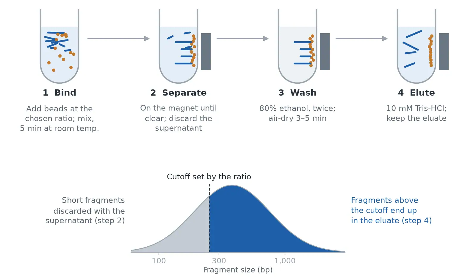
Bead volume equals sample volume multiplied by the ratio: a 50 µL sample at 1.8× takes 90 µL of bead suspension. Figure 1 shows the steps of a single-sided cleanup and which fragments each step removes.
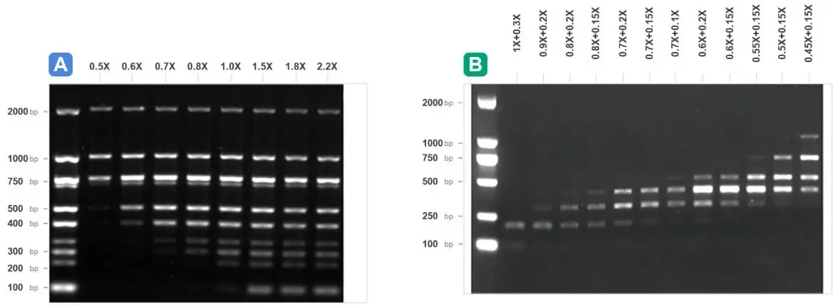
The cutoff is a slope, not a wall. In Figure 2A, fragments just below each listed cutoff are partly retained, while bands above it come through in full. Three conditions also move it: sample volumes below 50 µL, where pipetting errors become ratio errors (the BR3N401 manual recommends at least 50 µL); reaction buffers that already contain PEG, glycerol or high salt, which is why kit protocols state their own post-ligation ratios; and cold beads, so take BR3N401 out of 2–8 °C storage 30 minutes before use. Treat the table as a starting point and confirm it on your own ladder or library.
If your SOP was written for AMPure beads size selection, you can keep your ratios. BR3N401 uses an AMPure XP-like protocol, and the side-by-side gel on the product page shows closely matching band patterns at the same ratios.
Double-Sided Bead Based Size Selection for Narrow Insert Windows
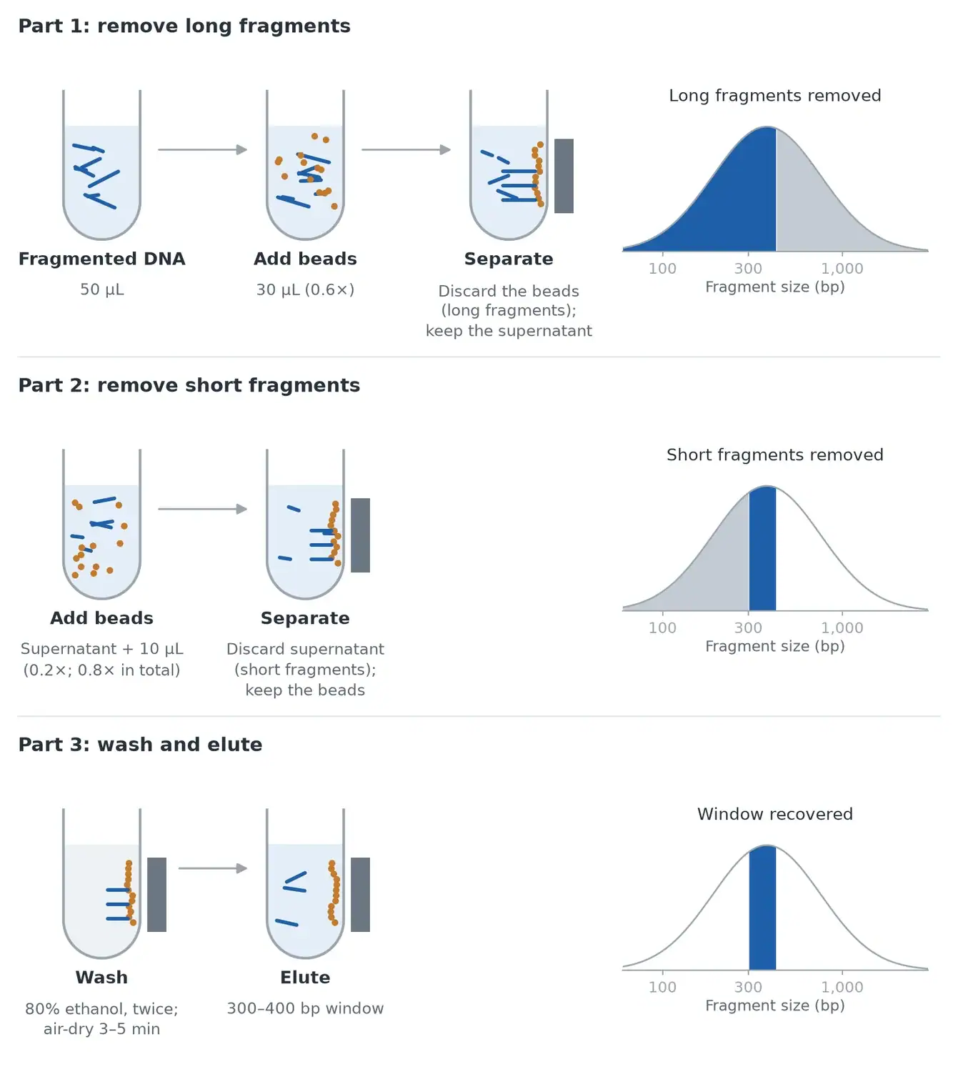
To keep a window instead of everything above a cutoff, add beads twice. The first addition is the larger one (0.6× against 0.2× in the example below): it binds the long fragments, so you discard those beads and keep the supernatant. The higher the first ratio, the lower the upper limit of the window. The second, smaller addition raises the total ratio so that the target window binds; you keep these beads and discard the supernatant, which carries the short fragments.
Both ratios are calculated on the original sample volume. For a 50 µL sample and a 300–400 bp window, add 30 µL of beads (0.6×), move the supernatant to a new tube, then add 10 µL (0.2×), bringing the total to 0.8×. Figure 3 follows this example step by step.
| Target window | First ratio | Second ratio | Total ratio |
|---|---|---|---|
| 100–200 bp | 1.0× | 0.3× | 1.3× |
| 200–300 bp | 0.7× | 0.2× | 0.9× |
| 300–400 bp | 0.6× | 0.2× | 0.8× |
| 400–500 bp | 0.5× | 0.15× | 0.65× |
| 400–700 bp | 0.45× | 0.15× | 0.6× |
In Figure 2B, as the first ratio drops from 1.0× to 0.45×, the retained bands move up from about 200 bp to the 400–750 bp range. A window costs yield, because everything outside it is discarded, so many workflows select after ligation and before PCR, where amplification can restore the mass.
Long-Read Libraries: Can SPRI Beads Enrich Fragments Above 5 kb?
Partly. A low bead ratio removes most short fragments, but a standard bead suspension does not give a sharp cutoff at 5 kb on its own.
The lowest single-sided ratio in Table 1, 0.5×, keeps fragments above about 1 kb. Below that, binding becomes harder to predict: the PEG concentration nears the point where even long DNA binds only in part, fewer beads are present to carry high-molecular-weight DNA, and viscous samples are hard to pipette accurately.
Long-read protocols work around this. Oxford Nanopore’s ligation sequencing protocol cleans up the adapter-ligated library at 0.4× and washes the beads with a Long Fragment Buffer instead of ethanol, which enriches fragments of 3 kb or longer.[4] Other workflows dilute the bead suspension in buffer to reach a lower PEG concentration with a pipettable volume, or use gel-based or precipitation-based kits built for high-molecular-weight DNA when a strict cutoff above 5 kb is needed. For background on the platforms, see our technical comparison of first-, second- and third-generation sequencing.
For long-read cleanups:
- Mix by flicking or slow pipetting with wide-bore tips to avoid shearing.
- Elute for 10 minutes; for very long DNA, Oxford Nanopore notes that 37 °C can improve recovery.[4]
- Do not over-dry: a cracked pellet releases long DNA poorly.
- Skip ethanol once nanopore sequencing adapters are attached, because it can denature the adapter’s motor protein.[4]
- Check sizes with pulsed-field electrophoresis or a genomic DNA capillary assay.
BR3N401’s recommended ratios run from 0.45× to 2.2×. To use it below 0.5× for long-read work, run a ratio test on your own high-molecular-weight DNA first. BIORI is also preparing dedicated products and protocols for long-fragment DNA selection, so keep an eye on our NGS range for updates.
FFPE and Low-Input Samples: Protect Recovery at Every Cleanup
Recovery losses multiply across steps. Three cleanups at 85% recovery keep 61% of the DNA; at 75% the same three keep 42%. For FFPE and low-input libraries, the number of cleanups and the recovery of each matter more than any other bead property.
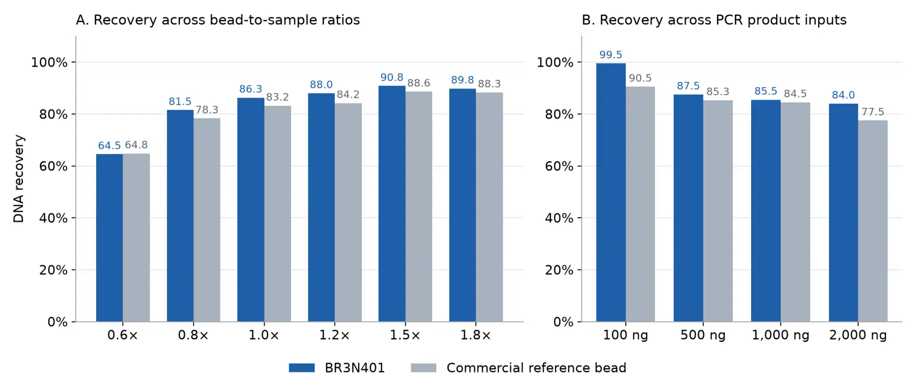
BIORI compared BR3N401 with a widely used commercial reference bead under the same protocol (Figure 4):
- From 0.8× to 1.8×, BR3N401 recovered more at every ratio (81.5–90.8% against 78.3–88.6%). At 0.6× both recovered about 65%, the yield traded for selectivity at a low ratio.
- At 100, 500, 1,000 and 2,000 ng input, BR3N401 recovered 99.5%, 87.5%, 85.5% and 84.0%, against 90.5%, 85.3%, 84.5% and 77.5%.
- In five replicates at 1.0×, BR3N401 averaged 83.6% (82.7–84.2%) against 77.1% (76.2–77.6%).
- In a separate test at 1.8× with 2,195 ng of amplified DNA, BR3N401 recovered 87.2–87.7%, in the top group of the five commercial bead products compared (72.4–88.3%).
FFPE DNA is already fragmented and carries damaged ends, so:
- Use 1.8× or higher after enzymatic steps so short, informative fragments are kept.
- Cut the number of cleanups. The AcuGenix™ Universal DNA Fragmentation Module (BR3N602) and the AcuGenix™ Universal DNA Library Prep Kit (BR3D201) run fragmentation, end repair and dA-tailing in one tube and remove three intermediate purifications; our article on enzyme-based library preparation explains the chemistry.
- Bring small reactions to at least 50 µL before adding beads, so the ratio stays accurate.
- Elute in a small volume of 10 mM Tris-HCl (pH 8.0) and incubate 2 minutes before returning the tube to the magnet.
Automating SPRI Cleanup: Settling Time and Bead Carry-Over
On a liquid handler, SPRI cleanup usually fails in one of three ways: beads settle in the reservoir, supernatant is removed before the beads have pelleted, or beads are drawn up with the eluate. Most fixes are settings.
- Mix the bead reservoir before each column. Beads settle in a trough within minutes, and a well that receives mostly buffer runs at a different effective ratio.
- Set magnet times by observation. The BR3N401 protocol binds for 5 minutes at room temperature, then waits on the magnet until the solution is clear. Clearing time depends on magnet strength, plate type and volume, so time it on your own deck with your largest volume and add a margin.
- Aspirate slowly with the tip offset from the pellet, and leave a few microliters behind rather than touch the beads.
- Wash twice with 200 µL of freshly prepared 80% ethanol for 30 s, then remove residual ethanol with a fine tip; ethanol in the eluate can inhibit the next enzyme.
- Air-dry for 3–5 minutes, until the pellet is no longer glossy, and check earlier on a heated deck.
- Keep beads out of the eluate. Trace carry-over causes trailing peaks on capillary electrophoresis traces.[3] Use a strong plate magnet, and if beads are visible, return the eluate to the magnet and transfer it again.
NGS DNA Clean Beads From BIORI
BIORI’s AcuGenix™ NGS DNA Clean Beads (BR3N401) are superparamagnetic SPRI beads in an optimized buffer, compatible with DNA and RNA library preparation kits from the major NGS suppliers. They follow an AMPure XP-like protocol, so existing ratios transfer without re-optimization, and they recovered 85% or more of input at standard cleanup ratios in head-to-head tests. They work manually or on automated liquid handling workstations, come in 1 mL, 5 mL, 60 mL and 450 mL packs with an instruction manual, COA and SDS, and are stored at 2–8 °C (do not freeze).
The beads pair with the BR3N602 fragmentation module, the BR3D201 library prep kit and the AcuGenix™ AmpSeq Library Amplification Mix (BR3M105). Browse all NGS modules or use the NGS library preparation product selection guide. BIORI manufactures under ISO 13485:2016 and ISO 9001:2015 certified quality systems and supplies bulk volumes for kit manufacturers.
FAQ
Do magnetic beads need to be completely dry before DNA elution?
No. Remove all of the ethanol, then air-dry for only 3–5 minutes, until the pellet is no longer glossy. A cracked pellet is over-dried and releases DNA less efficiently, especially long fragments; a wet pellet carries ethanol into the eluate, where it can inhibit the next enzymatic step.
How does magnetic bead separation work?
The chemistry and the magnet do separate jobs. PEG and salt in the bead buffer push DNA onto the carboxyl surface, and the bead-to-sample ratio decides which sizes bind. The magnet only collects the beads at the tube wall so the liquid can be removed. Washing and a low-salt elution buffer then release the purified DNA.
Which fraction should I keep after the first size-selection step?
Keep the supernatant. In double-sided selection the first bead addition, the larger of the two, binds the fragments that are too long, so those beads are discarded. After the second bead addition, keep the beads, which hold your target window, and discard the supernatant with the short fragments.
Can a published bead ratio predict recovery in my sample?
Only approximately. Ratio tables and recovery figures are measured on clean ladders or PCR products, and your sample’s buffer, size distribution, input mass and volume all shift the cutoff and the yield. In BIORI’s own data, recovery with the same bead ranged from 84.0% to 99.5% depending on input. Before locking an SOP, test two or three ratios on your own sample and check yield fluorometrically and size on a fragment analyzer.
What bead ratio removes adapter dimers?
A single 1.0× SPRI cleanup removes most adapter dimers, which typically run at about 120–150 bp, while keeping library fragments above about 300 bp. For longer-insert libraries, 0.8× removes them more strictly. If a dimer peak remains, repeat the cleanup at the same ratio rather than lowering it further.
Request a Sample
Tell us your library type, input range and automation platform through the AcuGenix™ NGS DNA Clean Beads page or our contact form, and we will follow up with a bead sample and ratio advice for your workflow.
NGS reagents are for research use only unless otherwise stated; bulk and custom formulations are supplied for further manufacturing use.
References
- DeAngelis MM, Wang DG, Hawkins TL. Solid-phase reversible immobilization for the isolation of PCR products. Nucleic Acids Res. 1995;23(22):4742–4743. doi:10.1093/nar/23.22.4742
- Lundin S, Stranneheim H, Pettersson E, Klevebring D, Lundeberg J. Increased throughput by parallelization of library preparation for massive sequencing. PLoS One. 2010;5(4):e10029. doi:10.1371/journal.pone.0010029
- BIORI. AcuGenix™ NGS DNA Clean Beads (BR3N401) Instruction Manual, V01. https://biori-en.com/wp-content/uploads/2026/06/Manual-BR3N401EN-AcuGenix-NGS-DNA-Clean-Beads-V01.pdf
- Oxford Nanopore Technologies. Ligation sequencing gDNA V14 (SQK-LSK114) protocol. https://nanoporetech.com/document/genomic-dna-by-ligation-sqk-lsk114