
RIPA Lysis Buffer (Medium)
Medium-lysis RIPA buffer with excellent cytoplasmic and good membrane and nuclear protein extraction for WB and IP.

Product overview
RIPA (Radio Immunoprecipitation Assay) Lysis Buffer is a conventional rapid cell and tissue lysis solution primarily used for extracting soluble proteins from animal cells and tissues. The protein samples obtained through lysis can be utilized for routine experiments such as Western Blot, immunoprecipitation (IP), and ELISA.
There are various formulations of RIPA lysis buffer, which can be broadly classified into strong, medium, and weak categories based on their lysis intensity. This product, RIPA Lysis Buffer (Medium), belongs to the Medium lysis category. Protein samples lysed with RIPA lysis buffer can be quantified using the BCA Protein Assay Kit (Biori#BR4C201) or Bradford Protein Assay Kit (Biori#BR4C202).
Choose the right RIPA strength
| BR4C101 Strong | BR4C102 Medium (this product) | BR4C103 Weak | |
|---|---|---|---|
| Effective lysis components | 1% Triton X-100, 0.75% sodium deoxycholate, 0.1% SDS | 1% NP-40, 0.5% sodium deoxycholate, 0.1% SDS | 1% NP-40, 0.05% sodium deoxycholate |
| Lysis strength | Strong | Medium | Weak |
| Membrane protein extraction | Excellent | Good | Average |
| Cytoplasmic protein extraction | Excellent | Excellent | Excellent |
| Nuclear protein extraction | Excellent | Good | Good |
| Main application | WB, IP | WB, IP | WB, IP, CO-IP |
All three strengths are also available together as the RIPA Lysis Buffer Set (BR4C104), 50 mL each.
Features and applications
- Medium lysis category: 1% NP-40, 0.5% sodium deoxycholate, 0.1% SDS.
- Take an appropriate amount of lysis buffer and add the corresponding protease/phosphatase inhibitor mixture (Biori#BR4C121/BR4C122/BR4C123) according to experimental requirements. Add the inhibitors at a ratio of 1:100 (V/V) within minutes prior to use.
- The protein concentration in the supernatant obtained after lysis with 100 µL of this product per 1 million animal cells is approximately 2–4 mg/mL, with variations observed depending on the cell type.
- For each 20 mg of cryopreserved mouse liver tissue lysed with 200 µL of this lysis buffer, the obtained supernatant exhibits a protein concentration of approximately 15–25 mg/mL.
- Western Blot (WB)
- Immunoprecipitation (IP)
- ELISA
- PAGE
Protocol
Notes
- This product is only for scientific research purposes, not for clinical diagnosis.
- The product must be thoroughly mixed before use, and all sample digestion steps should be performed on ice or at 4°C.
- A small amount of transparent gelatinous material frequently appears in the lysates of RIPA lysis buffer, which is a normal phenomenon and represents complexes formed with genomic DNA. If proteins tightly bound to genomic DNA are not to be detected, the supernatant can be directly obtained by centrifugation for subsequent experiments. For proteins requiring detection, the gelatinous material can be disrupted by ultrasonication followed by centrifugation to obtain the supernatant for further analysis. However, when detecting common transcription factors such as NF-κB or p53, ultrasonication is typically unnecessary for detection.
- For your safety and health, wear lab coats and disposable gloves during operation.
Cell sample lysis procedure
- Take an appropriate amount of lysis buffer and add the corresponding protease/phosphatase inhibitor mixture (Biori#BR4C121/BR4C122/BR4C123) according to experimental requirements. Add the inhibitors at a ratio of 1:100 (V/V) within minutes prior to use. Different concentrations of RIPA lysis buffer (Biori#BR4C104) can be selected based on experimental needs.
- For adherent cells: Remove the culture medium and wash once with PBS, normal saline, or serum-free medium (washing may be omitted if serum proteins do not interfere). Add lysate at a ratio of 150–250 µL per well in a 6-well plate, then vortex briefly to ensure thorough contact between the lysate and cells. Typically, cell lysis occurs after 1–2 seconds of lysate exposure to animal cells. For plant cells, lysis should be performed on ice for 2–10 minutes.
- For suspended cells: Collect cells by centrifugation, gently vortex or tap the tube bottom to maximize cell dispersion. Add lysis buffer at a ratio of 150–250 µL per well in a 6-well plate. Tap the tube bottom lightly to ensure complete cell lysis. After thorough lysis, no significant cell sediment should be observed. If the cell volume is substantial, aliquot the sample into tubes containing 500,000–1,000,000 cells per tube before proceeding with lysis.
Bacterial or yeast sample lysis procedure
- For 1 mL bacterial or yeast suspension, centrifuge at 10,000 rpm for 3–5 minutes and discard the supernatant. It is recommended to wash once with PBS buffer to thoroughly remove residual liquid, followed by gentle vortexing or pipetting at the tube bottom to maximize cell dispersion. Add 100–200 µL of lysis buffer, vortex gently or pipette at the tube bottom for homogenization, and lyse on ice for 2–10 minutes. For enhanced lysis efficiency, bacteria and yeast may be digested separately using lysozyme and lyticase, respectively, prior to lysis with the lysis buffer.
- After complete lysis, centrifuge at 12,000 rpm for 3–5 minutes, then collect the supernatant for subsequent procedures such as PAGE, Western blotting, and immunoprecipitation.
Note: Typically, 150 µL of lysis buffer is sufficient for each cell in a 6-well plate, based on the bacterial and yeast concentration in 1 mL of bacterial or yeast suspension. If the cell density is very high, the lysis buffer volume may be appropriately increased to 200–250 µL. The protein concentration in the supernatant obtained after lysis with 100 µL of this product per 1 million animal cells is approximately 2–4 mg/mL, with variations observed depending on the cell type.
Tissue sample lysis procedure
- Cut the tissue into small fragments. Homogenize using a glass homogenizer and grind with a tissue grinder until the sample is fully lysed. Alternatively, the tissue sample can be frozen and ground in liquid nitrogen, followed by addition of lysis buffer for lysis.
- Take an appropriate amount of lysis buffer and add an appropriate amount of protease and phosphatase inhibitor mixture (Biori#BR4C121/BR4C122/BR4C123) according to experimental requirements.
- Add the lysis buffer at a ratio of 150–250 µL per 20 mg of tissue (containing protease or phosphatase inhibitors). If lysis is incomplete, additional lysis buffer may be added appropriately. For high-concentration protein samples, the amount of lysis buffer can be reduced accordingly.
- After complete sample lysis, centrifuge at 12,000 rpm for 3–5 minutes and collect the supernatant for subsequent procedures such as PAGE, immunoblotting, and immunoprecipitation. For each 20 mg of cryopreserved mouse liver tissue lysed with 200 µL of this lysis buffer, the obtained supernatant exhibits a protein concentration of approximately 15–25 mg/mL. Protein concentrations may vary among tissue samples under different conditions following lysis.
Note: If the tissue sample itself is extremely fine, it can be appropriately cut and directly added to the lysis buffer for lysis, with thorough sample disruption achieved through vigorous vortexing. The supernatant obtained by centrifugation can be used for subsequent experiments. The advantage of direct lysis is its convenience and speed, eliminating the need for homogenizers or grinding equipment. However, the lysis efficiency is inferior to that of homogenization or grinding methods.
Components
| Components | BR4C102-01 |
|---|---|
| RIPA Lysis Buffer (Medium) | 100 mL |
Performance data
Comparison of Lysis Strength
Biori and Supplier N each used three RIPA Lysis Buffers of different strengths to extract proteins from 106 HEK293 cells. The resulting protein samples were then subjected to BCA concentration assay and Coomassie blue staining (SDS-PAGE/Coomassie staining).
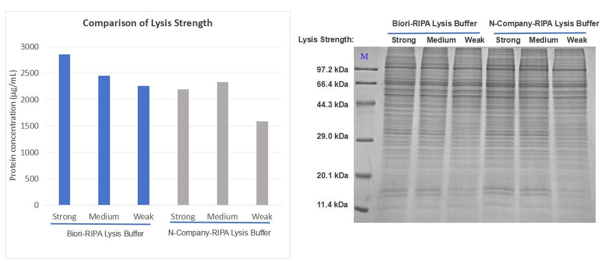
Conclusion: Biori RIPA buffers of three strengths showed a clearer concentration gradient than those of Supplier N, and all extracts showed intact protein bands on the Coomassie-stained gel.
Shipping and storage
| Condition | Recommendation |
|---|---|
| Storage | Store at 2-8°C. |
| Shipping | Ship at 2-8°C or under conditions that maintain 2-8°C. |
| Handling | For research use only. Review the MSDS before use. |
Downloads


Need this product lyophilized, in bulk, or in a custom format?
The feasibility assessment is free of charge. Send the assay brief and target format.